CHOLESTEROL OXIDASE from Microorganism
COO-331
Cholesterol:oxygen oxidoreductase(EC 1.1.3.6)

| Appearance: | Yellowish amorphous powder, lyophilized | |
|---|---|---|
| Activity: | GradeIII 12U/mg -solid or more |
|
| Contaminants: | Catalase ≤1.0×10⁻¹% Cholesterol esterase ≤1.0×10⁻²% |
|
| Stabilizers: | BSA, amino acids | |
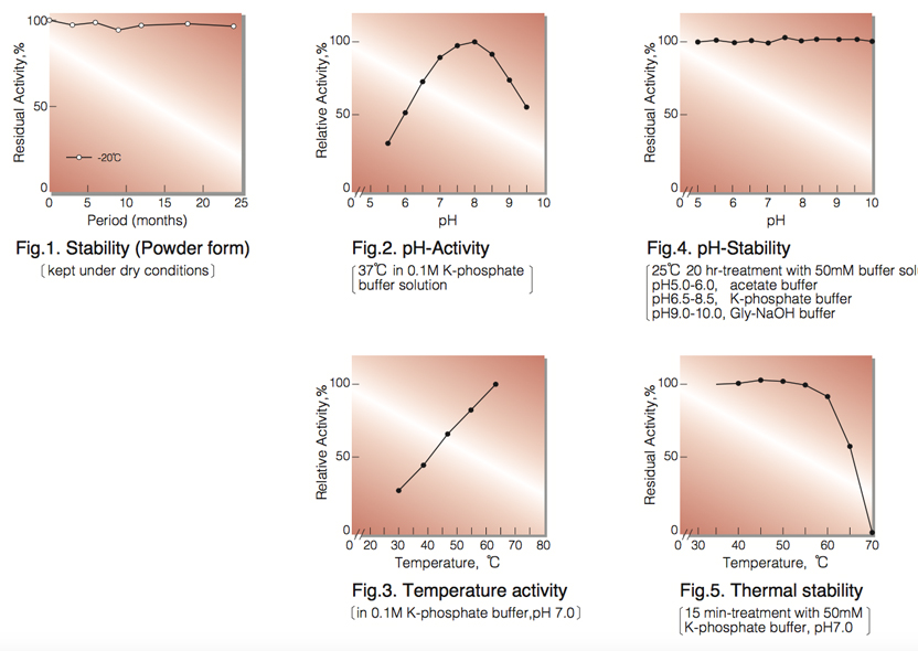
| Stability: | Stable at -20°C for at least One year (Fig.1) |
|---|---|
| Molecular weight: | approx. 55,000 (by gel-filtration) |
| Michaelis constants: | 3.0×10⁻⁵M(Cholesterol) |
| Inhibitors: | Ionic detergents, Hg⁺⁺ |
| Optimum pH: | 7.0-8.0(Fig.2) |
| Optimum temperature: | 60℃(Fig.3) |
| pH Stability: | pH 5.0-10.0 (25℃, 20hr)(Fig.4) |
| Thermal stability: | below 60℃ (pH 7.0, 15min)(Fig.5) |
| Substrate specificity: | (Table 1) |
| Effect of various chemicals: | (Table 2) |
APPLICATIONS
This enzyme is useful for enzymatic determination of cholesterol in serum when coupled with cholesterol esterase (COE-301, COE-311, COE-313) in clinical analysis.
ASSAY
Principle:
cholesterol oxidase
Cholesterol +O₂ ►Cholest-4-en-3-one+H₂O₂
peroxidase
2H₂O₂+4-Aminoantipyrine+Phenol ►Quioneimine dye+4H₂O
The appearance of quinoneimine dye formed when coupled with 4-aminoantipyrine and phenol is measured at 500nm by spectrophotometry.
Unit definition:
One unit causes the formation of one micromole of hydrogen peroxide (half a micromole of quinoneimine dye) per
minute under the conditions described below.
Method:
| A. 0.1M K-Phosphate buffer, pH 7.0 |
|
|---|---|
| B. Cholesterol solution: | To 5.0ml of Triton X-100 on a hot plate or in a water bath, add 500mg of cholesterol and mix with a stirring bar until cholesterol dissolves. Add 90ml of distilled water to the hot cholesterol-Triton X-100 solution by slowly pouring along a stirring bar. Stir and allow to boil for 30 to 60 seconds. The solution will be cloudy. Cool under running water with gentle agitation, the solution will turn clear. Add 4.0g of sodium cholate and dissolve. Fill up the solution to 100ml with distilled water. This solution is stable for about one week at room temperature. If it becomes cloudy, warm slightly while stirring until it clears. |
| C. 4-AA solution: | 1.76% (1.76g 4-aminoantipyrine/100ml of H₂O) |
| D. Phenol solution: | 6.0% (6.0g phenol/100ml of H₂O) |
| E. POD solution: | Horseradish peroxidase 15,000 purpurogallin units/100ml of buffer (A) |
| F. Enzyme diluent: | 20mM K-Phosphate buffer, pH 7.0 contg. 0.2% bovine serum albumin |
Procedure
| Concentration in assay mixture | |
|---|---|
| K-Phosphate buffer | 87 mM |
| Cholesterol | 0.89mM |
| 4-Aminoantipyrine | 1.4 mM |
| Phenol | 21 mM |
| Triton X-100 | 0.34 % |
| Sodium cholate | 64 mM |
| BSA | 33µg/ml |
| POD | 5.U/ml |
1. Prepare the following working solution (20 tests volume), immediately before use and store on ice in a brownish bottle.
51.0ml Buffer solution (A)
4.0ml Substrate solution (B)
1.0ml 4-AA solution (C)
2.0ml POD solution (E)
2. Pipette 2.9ml of working solution into a cuvette (d=1.0cm) and equilibrate at 37℃ for about 3 minutes. Add 0.1ml of Phenol solution (D), mix and keep at 37℃ for another 2 minutes.
3.Add 0.1ml of the enzyme solution* and mix with gentle inversion.
4. Record the increase in optical density at 500nm against water for 3 to 4 minutes in a spectrophotometer
thermostated at 37°C, and calculate the ΔOD per minute from the initial portion of the curve (ΔOD test).
At the same time, measure the blank rate (ΔOD blank) by using the same method as the test except that
the enzyme diluent is added instead of the enzyme solution.
* Dissolve the enzyme preparation in ice-cold enzyme diluent (F), and dilute to 0.1-0.3U/ml with the same buffer, and store on ice.
Calculation
Activity can be calculated by using the following formula :
ΔOD/min (ΔOD test−ΔOD blank ) × Vt × df
Volume activity (U/ml) = =ΔOD/min×4.499×df
13.78×1/2×1.0×Vs
Weight activity (U/mg) = (U/ml) × 1/C
- Vt
- : Total volume (3.1ml)
- Vs
- : Sample volume (0.1ml)
- 13.78
- : Millimolar extinction coefficient of quinoneimine dye under the assay conditions(㎠/micromole)
- 1/2
- : Factor based on the fact that one mole of H₂O₂ produces half a mole of quinoneimine dye
- 1.0
- : Light path length (cm)
- df
- : Dilution factor
- C
- : Enzyme concentration in dissolution (c mg/ml)
REFERENCES
- W.Richmond; Clin.Chem., 19, 1350 (1973).
- H.M.Flegg; Ann.Clin.Biochem., 10, 79 (1973).
- C.C Alain et al; Clin.Chem., 20, 470 (1974).
- P.N.Tarbutton and C.R.Gunter; Clin.Chem., 20, 724 (1974).
- S.Nomoto; Rinsho Kensa, 20, 688 (1976).
- K.Kameno et al; Jap.J.Clin.Path., 24, 650 (1976).
- Y.Nishiya et al; Protein Engng,7, 231 (1997)
| Substrate(0.1mM) | Relative activity(%) | Substrate(0.1mM) | Relative activity(%) |
|---|---|---|---|
| Cholesterol | 100.0 | Ergosterol | 44.0 |
| Pregnenolone | 60.0 | Lanosterol | 1.6 |
| ß-Cholestanol | 60.0 | Testosterone | 1.0 |
| ß-Sitosterol | 120.0 | Androsterone | 1.5 |
| Stigmasterol | 34.0 | Dehydroiso-androsterone | 15.0 |
| Chemical | Concn.(mM) | Residual activity(%) |
Chemical | Concn.(mM) | Residual activity(%) |
|---|---|---|---|---|---|
| None | − | 100 | NaF | 20 | 100 |
| Metal salt | 2.0 | NaN₃ | 20 | 100 | |
| MgCl₂ | 100 |
EDTA-2Na | 5.0 | 100 | |
| CaCl₂ | 100 | o-Phenanthroline | 2.0 | 100 | |
| Ba(OAc)₂ | 100 | α,α'-Dipyridyl | 1.0 | 100 | |
| FeCl₃ | 100 | Borate | 50 | 100 | |
| CoCl₂ | 100 | IAA | 2.0 | 100 | |
| MnCl₂ | 100 | NEM | 2.0 | 100 | |
| Zn(OAc)₂ | 100 | Hydroxylamine | 2.0 | 100 | |
| Cd(OAc)₂ | 100 | 2-Mercaptoethanol | 2.0 | 100 | |
| NiCl₂ | 100 | Triton X-100 | 0.10% | 100 | |
| CuSO₄ | 90 | Tween 20 | 0.10% | 94 | |
| Pb(OAc)₂ | 100 | Span 20 | 0.10% | 90 | |
| HgCl₂ | 0 | Na-cholate | 0.10% | 100 | |
| PCMB | 2.0 | 100 | SDS |
0.05% | 100 |
| MIA | 2.0 | 100 | DAC | 0.05% | 100 |
Ac, CH₃CO; PCMB, p-Chloromercuribenzoate; MIA, Monoiodoacetate; NEM, N-Ethylmaleimide; IAA, lodoacetamide; EDTA, Ethylenediamimetetraacetate;
SDS, Sodium dodecyl sulfate; DAC, Dimethylbenzylalkylammonium chloride.
To get a quote, contact us at info@toyobousa.com, or INQUIRY.